


Hi there! How can we help you today?

IVC Filter Placement
Inferior vena cava filter (IVC filter)
Inferior vena cava filter (IVC Filter)
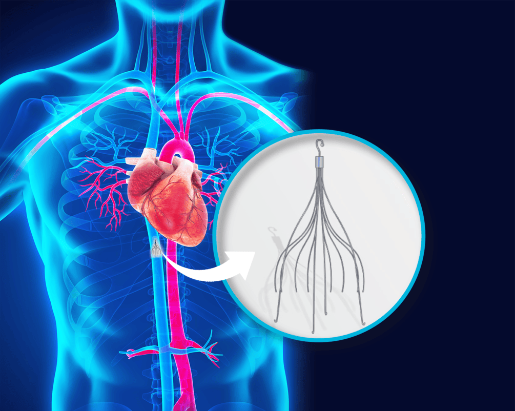
What is an inferior vena cava filter (IVC Filter)?
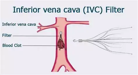
An inferior vena cava filter, or IVC Filter (Inferior Vena Cava Filter), is a specialized and important medical device placed in the large lower vein called the inferior vena cava (IVC) to help prevent pulmonary embolism (PE). The IVC is the largest vein in the human body and returns deoxygenated blood from the lower limbs and abdomen toward the heart. The main goal of placing this filter is to create a physical barrier that prevents blood clots formed in the lower limbs, usually the legs, from traveling to the lungs.
This specialized and sensitive procedure requires a high level of medical knowledge, experience, and skill and is professionally performed by Dr. Seyed Mohsen Jahromi Moghadam. His comprehensive and precise approach, from initial diagnosis and consultation to placement and follow-up, supports patient safety and peace of mind.
Structure and function of the IVC filter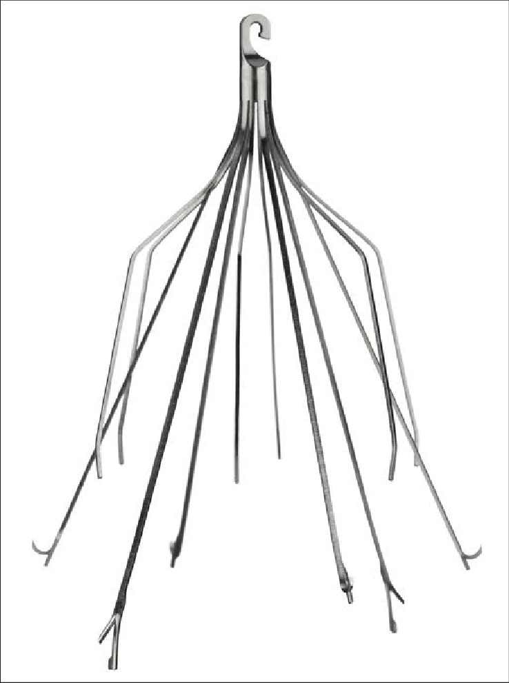
IVC filters are usually made from biocompatible metal alloys such as nitinol or stainless steel. They have an umbrella-like, basket-like, or mesh-like structure that opens after placement in the inferior vena cava and contacts the vein wall. Their design allows normal blood flow to continue while trapping large clots. Trapped clots are gradually broken down by the body's natural systems or become attached to the filter wall.
Indications for IVC filters: who needs this filter?
The decision to place an IVC filter is complex and is made individually after weighing risks and benefits. It is usually considered when other methods of preventing pulmonary embolism, especially anticoagulant medications, are ineffective or absolutely contraindicated.
Main indications for IVC filter placement include:
1. Patients with deep vein thrombosis (DVT) who have an absolute contraindication to anticoagulation or treatment failure with anticoagulants:
o Contraindication to anticoagulants: this includes patients with active and severe bleeding, such as brain bleeding, gastrointestinal bleeding, or bleeding after major surgery, in whom anticoagulation may be life-threatening. It also includes patients with inherited or acquired bleeding disorders.
o Failure of anticoagulant therapy: some patients develop progression of DVT or pulmonary embolism despite regular and adequate anticoagulation. In these cases, an IVC filter can provide an additional protective layer.
2. Patients who remain at high risk of pulmonary embolism despite anticoagulation:
o This group includes patients with a large clot burden, post-phlebitic syndrome, or other risk factors who remain at high risk for fatal PE despite anticoagulant medication. In these cases, the IVC filter may act as a bridge until the patient's condition stabilizes or anticoagulation becomes fully effective.
o High-risk patients with multiple trauma or prolonged hospitalization:
o Patients with severe trauma, spinal cord injury, or major orthopedic surgery are at very high risk of DVT and PE because of prolonged immobility, vascular injury, and severe inflammation. If anticoagulation cannot be used because of bleeding risk, prophylactic IVC filter placement may be considered.
o Patients with prolonged ICU admission or inability to move for different reasons may also fall into this category.
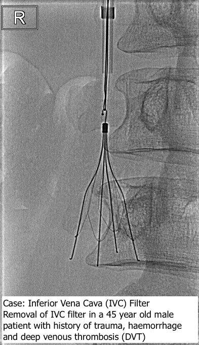
Dr. Seyed Mohsen Jahromi Moghadam evaluates the patient's medical history, current condition, and diagnostic tests such as lower limb venous Doppler ultrasound, pulmonary angiography, and coagulation tests to determine the best treatment approach. He explains the benefits and possible complications of filter placement to the patient and family so they can make an informed decision.
How an IVC filter is placed: a minimally invasive procedure
IVC filter placement is a specialized minimally invasive procedure usually performed as an outpatient procedure or with a short hospital stay. All steps, from preparation to placement and follow-up, are performed carefully by Dr. Seyed Mohsen Jahromi Moghadam.
Main steps include:
1. Patient preparation:
o Before the procedure, the patient undergoes a complete physical examination, and medical history and medications are reviewed.
o Blood tests, including complete blood count, coagulation tests, and electrolytes, are performed.
o The patient usually fasts for several hours before the procedure.
o The catheter entry site, usually the groin or neck, is sterilized and prepared.
2. Local anesthesia:
o Unlike many major surgeries, general anesthesia is not required.
o The physician injects local anesthetic such as lidocaine into the groin or neck area. The patient remains awake but does not feel pain. Mild sedation may be used if needed.
3. Venous access:
o After anesthesia, Dr. Jahromi Moghadam accesses a large vein, usually the femoral vein in the groin or the internal jugular vein in the neck, using a fine needle and the Seldinger technique.
o A thin guidewire is inserted through the needle, and the needle is removed.
o A sheath, about 5 to 7 mm in diameter, is inserted over the wire to create a safe route for the filter.
4. Image guidance (fluoroscopy):
o Live X-ray imaging allows the physician to see the guidewire, sheath, and filter in real time.
o Contrast material may be injected to visualize the IVC and nearby vessels and identify the exact site for filter placement.
o The ideal position is usually below the renal veins so kidney blood flow is not obstructed while clots from the lower limbs can be trapped.
5. Filter deployment:
The compressed IVC filter inside a delivery catheter is advanced through the sheath.
o The catheter is guided under fluoroscopy to the correct position in the IVC.
o Once correctly positioned, the filter is slowly released, opens inside the vein, and contacts the wall. Its shape traps clots while normal blood flow continues.
6. Confirming position and removing instruments:
o After release, the physician confirms proper position and complete opening using fluoroscopy or contrast injection.
o The sheath and guidewire are then removed.
o Pressure is applied to the puncture site to stop bleeding, followed by a pressure dressing.
Timing and discharge:
The whole filter placement process usually takes less than one hour. Many patients are discharged the same day after a few hours of rest and observation. Some patients with more complex conditions may stay overnight.
Dr. Seyed Mohsen Jahromi Moghadam personally performs and follows all stages of this sensitive procedure, ensuring maximum safety and effectiveness and providing detailed post-procedure care instructions.
Types of IVC filters: permanent and retrievable
IVC filters are divided into two main categories. The choice depends on the patient's clinical condition, expected duration of PE risk, and the physician's judgment.
1. Permanent IVC filters:
o These filters are designed to remain in the IVC permanently. They are made of materials safe for long-term use and usually have struts that secure them to the vein wall.
o They are used for patients with long-term or permanent PE risk or lifelong contraindication to anticoagulation.
o Advantage: no second procedure is needed for removal.
o Limitation: long-term complications such as venous obstruction or filter fracture may be harder to manage.
2. Retrievable or temporary IVC filters:
o These filters are designed to be removed after the risk of PE resolves or when the patient can safely receive anticoagulation. They often have a hook or loop that allows retrieval.
o They are used when PE risk is temporary, such as after major surgery, during active bleeding that is expected to resolve, in trauma patients, or in some pregnant patients at risk of DVT/PE.
o Advantage: removal can reduce long-term filter-related risks such as IVC obstruction, fracture, or vessel perforation.
o Limitation: removal requires a second procedure, and if not removed on time they may behave like permanent filters.
Dr. Jahromi Moghadam evaluates each patient carefully and recommends the most suitable filter type based on expected DVT/PE risk duration, ability to use anticoagulants, and overall health. He also manages the timing of retrievable filter removal.
Benefits and possible complications: a comprehensive assessment
Like any medical procedure, IVC filter placement has benefits and possible complications that must be evaluated carefully. Dr. Jahromi Moghadam explains all aspects transparently so the patient and family can make an informed decision.
Benefits:
The main and most important benefit is a significant reduction in the risk of pulmonary embolism in selected high-risk patients, especially when anticoagulants are contraindicated, ineffective, or when prophylaxis is needed in very high-risk trauma or immobilized patients.
By preventing pulmonary embolism, an IVC filter can directly save life and prevent serious or fatal complications such as cardiopulmonary failure, stroke-like events, or death.
Possible complications:
Although IVC filter placement is relatively safe, possible complications exist. Dr. Jahromi Moghadam uses precise protocols and modern equipment to minimize these risks.
Short-term complications, rare:
• Bleeding or bruising at the catheter entry site
• Infection at the entry site
• Vessel injury during placement
• Allergic reaction to contrast material
• Contrast-related kidney injury in patients with kidney dysfunction
Long-term complications, rare:
• Clot formation inside the filter, or filter thrombosis, which can cause IVC obstruction, leg swelling, pain, and chronic discomfort
• Filter migration toward the heart or lungs
• Filter fracture, with small pieces traveling in the bloodstream
• Vena cava penetration or perforation into adjacent structures
• Failed retrieval of a retrievable filter because of tissue ingrowth or filter deformation
• Possible increased future DVT risk, especially with long-term filters
Serious IVC filter complications are uncommon, but individualized risk-benefit assessment and performance by experienced specialists are essential.
Care and follow-up: ensuring proper function and patient safety
After IVC filter placement, careful and regular follow-up is important to ensure proper filter function and overall patient health. Dr. Jahromi Moghadam creates individualized follow-up plans that may include:
1. Immediate post-procedure care:
o Monitoring the catheter entry site for bleeding, bruising, or infection.
o Several hours of rest after the procedure.
o Adequate fluids to help eliminate contrast material, and simple pain medication if needed.
2. Regular follow-up visits:
o An early follow-up visit within the first few weeks to assess general status and the access site.
o Periodic imaging, such as lower limb venous Doppler ultrasound, plain X-ray, CT scan, or intravascular ultrasound, depending on filter type and patient condition.
o Assessment and adjustment of anticoagulant medication when indicated.
3. Management of retrievable filters:
o For patients with retrievable filters, a removal plan is made. Removal is often recommended after the risk has resolved, commonly between 3 weeks and 6 months, to reduce long-term complications.
o Filter retrieval is also performed under local anesthesia and image guidance and is usually shorter and simpler than placement.
4. Monitoring for warning symptoms:
o Patients are educated to seek care for new severe leg swelling, pain or redness, chest pain, shortness of breath, coughing blood, fever, chills, or unusual abdominal or back pain.
The role of Dr. Seyed Mohsen Jahromi Moghadam in care and follow-up:
As the physician responsible for IVC filter placement, Dr. Jahromi Moghadam plays a central role in all aspects of care and follow-up. He follows standard protocols, decides when imaging is needed, interprets the results, plans retrieval of retrievable filters, promptly manages any complications, and remains available for patient questions and consultation.
________________________________________
For more information or consultation about IVC filter placement, you can contact Delban Specialized Clinic and Dr. Seyed Mohsen Jahromi Moghadam.
05132042 - 09003204200