


Hi there! How can we help you today?

Lower Limb Arterial Angiography
Peripheral artery disease (PAD)
Leg arterial angiography and stenting
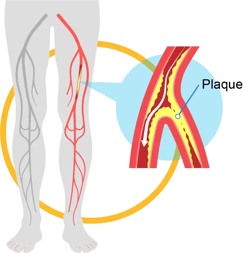
Introduction to peripheral artery disease (PAD)
Peripheral artery disease, or PAD, refers to reduced blood flow to the lower limbs due to narrowing or blockage of the arteries of the thigh, leg, and foot. This disorder is mainly caused by atherosclerosis, which reduces the delivery of oxygen and nutrients to tissues. PAD significantly lowers quality of life and can lead to pain while walking, chronic wounds, and, if advanced, embolic events or vascular insufficiency. Early diagnosis and timely management through a comprehensive combination of healthy lifestyle changes, medications, and endovascular interventions help improve medium-term and long-term patient outcomes.
Symptoms, causes, and the importance of early diagnosis
- Common symptoms: pain or cramping in the calf while walking that improves with rest (claudication), tingling or itching, coldness or numbness of the feet, foot ulcers or gangrene in advanced stages, skin discoloration, and thickened nails.
- Main causes: atherosclerotic disease, high blood pressure, diabetes mellitus, dyslipidemia, smoking, family history, physical inactivity, and inflammatory factors.
- Importance of early diagnosis: preventing disease progression, avoiding severe complications such as diabetic foot ulcers, reducing the risk of hospitalization, and preserving the patient's walking ability.
What is leg arterial angiography and when is it performed?
- Definition: Leg arterial angiography is an invasive imaging procedure in which contrast material is injected and images are taken of the leg arteries to show the exact location and severity of narrowing or blockage.
When it is performed: It is done when PAD symptoms suggest a high likelihood of arterial narrowing, when noninvasive imaging such as Doppler ultrasound or CT/MRI angiography does not provide sufficient information, or to plan endovascular treatment such as angioplasty or stenting. It may also be performed after noninvasive assessment to determine whether intervention is needed or to complete the diagnostic process.
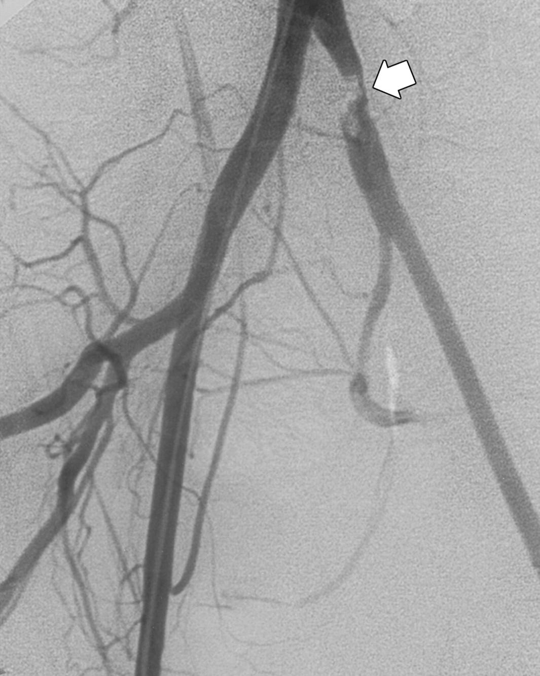
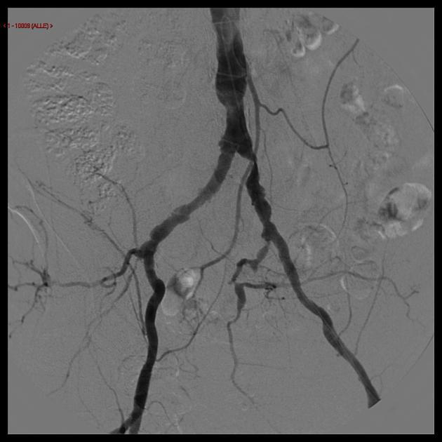
Steps of leg arterial angiography and stenting
- Preparation and initial assessment:
- Medical history and medication review, blood tests, blood pressure assessment, and coagulation profile evaluation.
- Patient education about the purpose, risks, and benefits, followed by obtaining informed consent.
- Discontinuation of anticoagulant medications if needed according to the physician's instructions and in coordination with the medical team.
- Entering the angiography suite and anesthesia:
- Local anesthesia in the arm or leg area, and sometimes light sedation for patient comfort.
- Use of full sterile technique and a specialized vascular team.
- Catheter insertion and contrast injection:
- Advancing the catheter through the vessels toward the affected area and injecting contrast material to display the vascular anatomy on fluoroscopic or CT images.
- Obtaining detailed images of the full length of the leg vessels and foot arteries.
- Precise diagnosis and decision-making about intervention:
- Determining the location and severity of narrowing or blockage, assessing whether stenting is feasible, and considering other techniques such as balloon angioplasty, atherectomy, or vascular grafting.
- Leg arterial stenting (if needed):
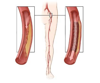
- Opening the vessel with a balloon and placing a metal stent to keep the artery open over the long term and maintain blood flow.
- Adjusting balloon pressure and reassessing blood flow after stent placement with repeat imaging.
- Completion and return to normal status:
- Dressing the puncture site and transferring the patient to the ward for monitoring.
Providing post-procedure care and medication instructions to prevent thrombosis and recurrent narrowing.
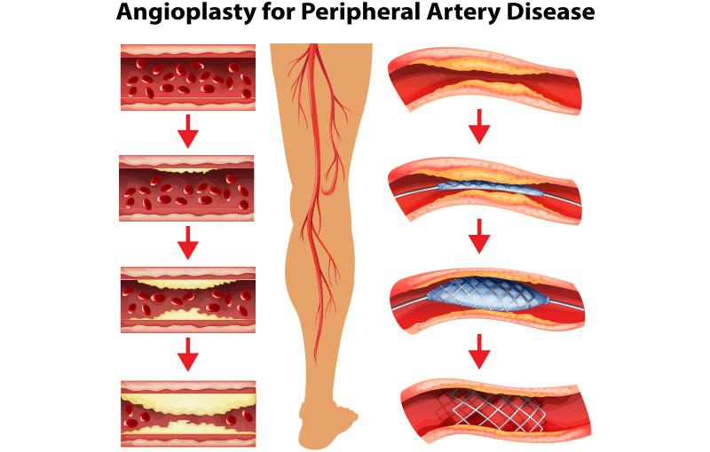
Benefits and treatment outcomes
- Significant improvement in arterial blood flow to the legs and reduction of pain while walking.
- Improved walking capacity, quality of life, and lower limb function.
- Reduced risk of foot ulcers and the need for extensive open surgeries.
- Stenting and other vascular interventions can help maintain long-term vessel patency.
- Treatment outcomes strongly depend on appropriate patient selection and risk factors such as diabetes or nicotine use.
Possible complications and frequency
- Short-term complications: local pain, bruising, bleeding at the catheter entry site, sensitivity to contrast material, and superficial infections.
- Rare but more serious complications: seizure, vascular injury, embolism or clot formation, and severe reaction to contrast material.
- Frequency of complications: With an experienced team, careful monitoring, and safe techniques, serious complications are relatively uncommon; however, risks must be explained to the patient in each case.
Care before and after angiography
Before angiography:
- Avoiding food and certain medications according to the medical team's instructions.
- Coordination with the nursing team regarding medications and the pre-procedure regimen.
- Stopping or reducing tobacco use to improve treatment outcomes.
After angiography:
- Resting in the ward for a defined period, with blood pressure and blood status monitoring.
- Monitoring the catheter entry site and avoiding excessive pressure on the area.
- Taking anticoagulant or antiplatelet medications as prescribed by the physician.
Regular follow-up, pain control, and education about warning signs such as bleeding or infection.
ABI test
Definition of ABI
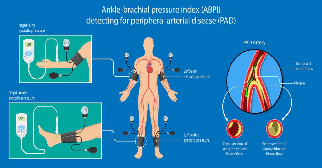
- ABI, or ankle-brachial index, is the ratio of ankle pressure to arm pressure and is a simple, noninvasive index for assessing lower limb blood flow.
- Values within or above the normal range indicate healthy vessels, while values below normal suggest narrowing or blockage in the leg arteries.
How the ABI test is performed, in simple scientific language
- Performed noninvasively: systolic pressure is measured in both legs and both arms using specialized pressure cuffs.
- ABI is calculated for each leg by dividing the ankle pressure by the arm pressure.
- The measurement is repeated in both legs for comparison.
Interpretation of ABI values (normal, mild, moderate, severe)
- Normal: 1.0 to 1.4
- Mild reduction: 0.91 to 0.99
- Moderate to severe reduction: 0.41 to 0.90
- Very severe disease or complete occlusion: less than 0.40
- Values above 1.4 may indicate noncompressible arteries or measurement interference.
Diseases detectable with ABI
- PAD of varying severity, from moderate to severe obstruction.
- Associated vascular disorders such as impaired arterial function or arterial insufficiency.
Importance of ABI in treatment decision-making
- ABI is used as an initial screening tool and severity assessment method to support better decisions about interventions such as stenting or noninvasive treatment.
- When combined with other diagnostic tools such as Doppler ultrasound and imaging, ABI provides a more accurate estimate of vascular status.
Benefits and treatment outcomes
- Fast and inexpensive diagnosis of PAD.
- Precise guidance in selecting noninvasive treatments or interventions such as leg angiography and stenting.
- Continuous ABI monitoring to follow disease progression or improvement.
Possible complications and frequency
- The ABI test has no serious complications and is generally safe.
- Temporary pain or bruising after cuff pressure on the leg may occur but is usually mild.
- Complications of ABI are very rare and are mostly related to superficial discomfort.
ABI testing, diagnosis, and treatment of peripheral artery disease at Delban Advanced Cardiovascular Clinic
- Delban Clinic diagnoses and treats PAD with a team of cardiovascular specialists and in-house imaging teams.
- Services include ABI, vascular Doppler ultrasound, leg arterial angiography, stenting, and modern vascular interventions.
- High medical standards are followed to maintain quality, safety, and patient satisfaction.
- Through careful evaluation and treatment planning, Delban Clinic helps improve lower limb function and patients' quality of life.
Final summary
Leg arterial angiography and stenting, as a central component of interventional PAD treatment, can improve quality of life, reduce pain, and preserve mobility when supported by accurate diagnosis and timely intervention. The ABI test is a key noninvasive tool for screening and treatment decision-making, and at Delban Advanced Cardiovascular Clinic it is provided continuously with experienced care, early diagnosis, and effective treatment. Our goal is to deliver the highest quality medical services with the lowest possible risk for patients.